Patent ductus arteriosus (PDA) is a heart problem that occurs soon after birth in some babies. In PDA, there is an abnormal circulation of blood between two of the major arteries near the heart. Before birth, the two major arteries-the aorta and the pulmonary artery-are normally connected by a blood vessel called the ductus arteriosus, which is an essential part of the fetal circulation.
After birth, the vessel is supposed to close within a few days as part of the normal changes occurring in the baby's circulation. In some babies, however, the ductus arteriosus remains open (patent). This opening allows blood to flow directly from the aorta into the pulmonary artery, which can put a strain on the heart and increase the blood pressure in the lung arteries.
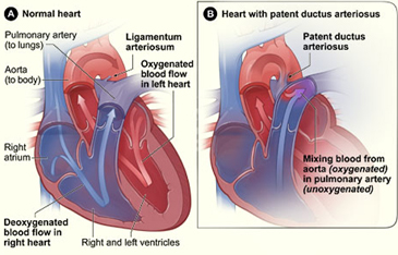
Figure A shows the normal anatomy and blood flow of the interior of the heart. Figure B shows a heart with a patent ductus arteriosus. The defect connects the aorta with the pulmonary artery, allowing oxygen-rich blood from the aorta to mix with oxygen-poor blood in the pulmonary artery.
A PDA is a type of congenital heart defect. A congenital heart defect is any type of heart problem that is present at birth.
If your baby has a PDA, but has an otherwise normal heart, the PDA may shrink and go away completely, or it may need to be treated to close it. But, if your baby is born with certain types of heart defects that decrease blood flow from the heart to the lungs or the body, medicine may be given to keep the ductus arteriosus open to maintain blood flow and oxygen levels until corrective surgery for the heart defect(s) can be performed.
About 3,000 infants are diagnosed with PDA each year in the United States. It is more common in premature infants (babies born too early) but does occur in full-term infants. Premature babies with PDA are more vulnerable to its effects. PDA is twice as common in girls as in boys.
The next section, How the Heart Works, provides a more detailed description of a heart with a PDA compared to a normal heart. See that section for a more detailed description of the anatomy and circulation of a normal heart.
How the Heart Works
Your child's heart is a muscle about the size of his or her fist. It works like a pump and beats 100,000 times a day.
The heart has two sides, separated by an inner wall called the septum. The right side of the heart pumps blood to the lungs to pick up oxygen. Then, oxygen-rich blood returns from the lungs to the left side of the heart, and the left side pumps it to the body.
The heart has four chambers and four valves and is connected to various blood vessels. Veins are the blood vessels that carry blood from the body to the heart. Arteries are the blood vessels that carry blood away from the heart to the body.
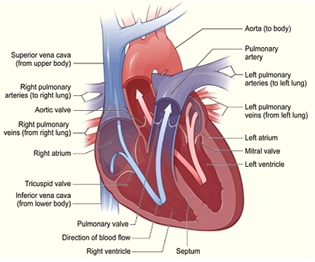
The illustration shows a cross-section of a healthy heart and its inside structures. The blue arrow shows the direction in which oxygen-poor blood flows from the body to the lungs. The red arrow shows the direction in which oxygen-rich blood flows from the lungs to the rest of the body.
Heart Chambers
The heart has four chambers or "rooms."
The atria (AY-tree-uh) are the two upper chambers that collect blood as it comes into the heart.
The ventricles (VEN-trih-kuls) are the two lower chambers that pump blood out of the heart to the lungs or other parts of the body.
Heart Valves
Four valves control the flow of blood from the atria to the ventricles and from the ventricles into the two large arteries connected to the heart.
The tricuspid (tri-CUSS-pid) valve is in the right side of the heart, between the right atrium and the right ventricle.
The pulmonary (PULL-mun-ary) valve is in the right side of the heart, between the right ventricle and the entrance to the pulmonary artery, which carries blood to the lungs.
The mitral (MI-trul) valve is in the left side of the heart, between the left atrium and the left ventricle.
The aortic (ay-OR-tik) valve is in the left side of the heart, between the left ventricle and the entrance to the aorta, the artery that carries blood to the body.
Valves are like doors that open and close. They open to allow blood to flow through to the next chamber or to one of the arteries, and then they shut to keep blood from flowing backward.
When the heart's valves open and close, they make a "lub-DUB" sound that a doctor can hear using a stethoscope.
The first sound-the "lub"-is made by the mitral and tricuspid valves closing at the beginning of systole (SIS-toe-lee). Systole is when the ventricles contract, or squeeze, and pump blood out of the heart.
The second sound-the "DUB"-is made by the aortic and pulmonary valves closing at beginning of diastole (di-AS-toe-lee). Diastole is when the ventricles relax and fill with blood pumped into them by the atria.
Arteries
The arteries are major blood vessels connected to your heart.
The pulmonary artery carries blood pumped from the right side of the heart to the lungs to pick up a fresh supply of oxygen.
The aorta is the main artery that carries oxygen-rich blood pumped from the left side of the heart out to the body.
The coronary arteries are the other important arteries attached to the heart. They carry oxygen-rich blood from the aorta to the heart muscle, which must have its own blood supply to function.
Veins
The veins are also major blood vessels connected to your heart.
The pulmonary veins carry oxygen-rich blood from the lungs to the left side of the heart so it can be pumped out to the body.
The vena cava is a large vein that carries oxygen-poor blood from the body back to the heart.
For more information on how a healthy heart works, see the Diseases and Conditions Index article on How the Heart Works. This article contains animations that show how your heart pumps blood and how your heart's electrical system works.
The Heart With Patent Ductus Arteriosus
The ductus arteriosus is a blood vessel that connects a baby's aorta and pulmonary artery while the baby is in the womb. This connection allows blood to be pumped from the right side of the heart straight to the aorta without stopping at the lungs for oxygen. In the womb, only a small amount of a baby's blood needs to go to the lungs because the baby gets oxygen from the mother's bloodstream. The baby's pulmonary artery, which carries blood to the lungs, is not needed at this time.
After birth, the baby is no longer connected to the mother's bloodstream. The baby's blood must now go to his or her own lungs to get oxygen. Normally, as the baby begins to breathe on his or her own, the pulmonary artery opens to allow blood into the lungs, and the ductus arteriosus closes. Once the ductus arteriosus closes, blood leaving the right side of the heart no longer goes straight to the aorta. First, it goes through the pulmonary arteries and stops at the lungs to pick up oxygen. Then, blood carrying oxygen returns to the left side of the heart and is pumped out to the rest of the body.
If the ductus arteriosus does not close after birth as it should, it is called a patent ductus arteriosus (PDA). A PDA allows blood to flow directly from the aorta into the pulmonary artery and from there to the lungs. This extra amount of blood flowing into the lungs strains the heart and increases the blood pressure in the arteries of the lungs.
Effects of patent ductus arteriosus
Normal birth-weight infants. The larger a PDA is, the greater the amount of extra blood that passes through the lungs. A small PDA might not cause any problems, whereas a larger PDA is likely to cause problems.
PDA can increase the risk of bacterial endocarditis. Bacterial endocarditis is an infection of the lining of the heart, valves, or arteries. In the case of PDA, the increased flow of blood can irritate the lining of the pulmonary artery where the PDA connects. This irritation makes it easier for bacteria in the bloodstream to collect and grow there.
A large PDA that is allowed to remain open for an extended period of time can cause the heart to enlarge and to have to work harder. Also, fluid can build up in the lungs.
Premature infants. For premature infants (babies born too early), PDA can be more serious than in normal-weight babies. Preemies with PDA are more likely to have damage to their lungs from the extra blood flowing through the PDA. Preemies with PDA may need to be on a ventilator to help them with their breathing.
The increased flow of blood through the lungs also can reduce blood flow to the rest of the body. This can damage other organs, especially the intestines and kidneys.
What Causes Patent Ductus Arteriosus ?
The cause of patent ductus arteriosus (PDA) is not known.
Genetics may play a role. A defect in one or more genes could prevent the ductus arteriosus from closing normally after birth.
PDA is more common in : -
Premature infants (babies born too early)
Infants with genetic abnormalities such as Down syndrome
Infants whose mother had German measles (rubella) during pregnancy
What Are the Signs and Symptoms of Patent Ductus Arteriosus ?
A heart murmur may be the only sign that a baby has patent ductus arteriosus (PDA). A heart murmur is an extra or unusual sound heard during the heartbeat.Some infants may develop signs or symptoms of volume overload on the heart and excess blood flow in the lungs.
Signs and symptoms may include : -
Fast breathing, working hard to breathe, or shortness of breath, or in the case of a premature infant, need for increased oxygen or ventilatory support
Poor feeding and poor weight gain
Tiring easily
Sweating with exertion (such as while feeding)
How Is Patent Ductus Arteriosus Diagnosed ?
In full-term infants, a patent ductus arteriosus (PDA) usually is first suspected when the baby's doctor hears a heart murmur during a regular checkup. If a PDA is large, the infant may also develop symptoms of volume overload and increased blood flow to the lungs. When a PDA is small, it may not be diagnosed until later in childhood. Once a PDA is suspected, a consultation with a pediatric cardiologist will be arranged. A pediatric cardiologist is a doctor who specializes in diagnosing and treating heart problems in children.
In premature babies (babies born too early) with PDA, the physical signs that are seen in full-term babies, such as heart murmur, may not be present. Doctors may suspect a PDA in premature babies who develop breathing difficulties soon after birth. Doctors use tests such as echocardiography to look for PDA in premature babies with breathing problems.
Tests
Two painless tests are used to diagnose a PDA : -
Echocardiogram : - This test, which is harmless and painless, uses sound waves to create a moving picture of your baby's heart. During an echocardiogram, reflected sound waves outline the heart's structure completely. The test allows the doctor to clearly see any problem with the way the heart is formed or the way it's working.
An echocardiogram is the most important test available to your baby's cardiologist to both diagnose a heart problem and follow the problem over time. In babies with PDA, the echocardiogram shows how big the ductus is and how well the heart is responding to it. When medical treatments are used to try to close a ductus in premature babies, echocardiograms are used to see how well the treatment is working.
EKG (electrocardiogram) : - This test records the electrical activity in the heart.
In the case of a PDA, it can show : -
Enlargement of the heart chambers
Other subtle changes that can suggest the presence of a PDA
How Is Patent Ductus Arteriosus Treated ?
The goal of treatment is to close the patent ductus arteriosus (PDA) to prevent complications and reverse the effects of increased blood volume.
Small PDAs often close without treatment.
For full-term infants, treatment is needed if the child's PDA : -
Is large
Is causing the child to have health problems
Does not close on its own by the time the child is 1-2 years old
For premature infants (babies born too early), treatment is needed if the PDA is causing increased respiratory distress and heart problems.
Your child's doctor will discuss the treatment options and your family's preferences regarding treatment decisions.
Medicines : -
Medicines can be given to help close a PDA.
Indomethacin (in-doh-METH-ah-sin) is a drug that helps close a PDA in premature infants. It does not usually work in full-term infants. It works by stimulating the PDA to constrict or tighten, closing the connection.
Ibuprofen (EYE-boo-pro-fen) is a medicine in the same family as indomethacin. It is also used frequently to close a PDA in premature infants.
If a PDA is small and the decision is made not to treat it right away, antibiotics may be prescribed to prevent endocarditis.
Catheter-based procedures : -
Catheters are thin, flexible tubes used in a procedure called cardiac catheterization (KATH-e-ter-i-ZA-shun). Catheter-based procedures are often used to close PDAs in infants or children who are large enough to have the procedure. Your child's doctor may refer to the procedure as "transcatheter device closure." The procedure is sometimes done on small PDAs to prevent the risk of bacterial endocarditis.
During the procedure, your child will be sedated or given general anesthesia so he or she will sleep and not feel any discomfort. The doctor will place a catheter in a large blood vessel in the upper thigh (groin) and guide it to your child's heart.
A small metal coil or other blocking device is passed up through the catheter and placed in the ductus arteriosus to block blood flow through the vessel.
Catheter-based procedures : -
Do not require the child's chest to be opened
Let the child recover quickly
Closing a PDA using a catheter is often done on an outpatient basis. You will most likely be able to take your child home the same day the procedure is done.
Complications of catheter-based procedures are rare and short term. They can include bleeding, infection, and movement of the blocking device from where it was placed.
Surgery : -
Surgery for PDA may be performed when : -
A premature or full-term infant develops health problems from the PDA and is too small to have a catheter-based procedure
A PDA is not successfully closed by a catheter-based procedure
Surgery is planned for treatment of related congenital heart defects
Surgery often is not performed until after 6 months of age in infants who do not have health problems from the PDA. Doctors sometime perform surgery on small PDAs to prevent the risk of bacterial endocarditis.
The operation is done under general anesthesia so that your child will sleep and not feel any pain.
The surgeon will : -
Make a small cut between your child's ribs to reach the PDA
Close the PDA with stitches or clips
Complications of the surgery are rare and usually short term. They can include hoarseness, a paralyzed diaphragm, infection, bleeding, or fluid buildup around the lungs.
After surgery. After surgery, your child will spend a few days in the hospital. Most children go home 2 days after surgery. While in the hospital, your child will be given medicines to reduce pain or anxiety. The doctors and nurses at the hospital will teach you how to care for your child at home
They will talk to you about : -
Limits on activity for your child while he or she recovers
Followup appointments with your child's doctors
How to give your child medicines at home, if needed
When your child goes home after surgery, you can expect that he or she will feel fairly comfortable, although there may be some pain temporarily.
Your child should begin to eat better and gain weight quickly. Within a few weeks, your child should be fully recovered and able to participate in normal activities.
Long-term complications from surgical treatment are rare. They can include narrowing of the aorta, incomplete closure of the ductus arteriosus, and reopening of the ductus arteriosus.
The list of of world class heart hospitals in India is as follows : -
For more information, medical assessment and medical quote
send your detailed medical history and medical reports
as email attachment to
Email : - info@wecareindia.com
Call: +91 9029304141 (10 am. To 8 pm. IST)
(Only for international patients seeking treatment in India)
For a detailed evaluation send patient’s medical reports / X rays / doctors notes to info@wecareindia.com
Patient Storys
Successful heart surgery at We Care India partner hospital allows Robert Clarke to live a normal life despite a rare genetic disorder We Care india helped Robert find best super specialised surgeon for his rare conditions.