We Care partner hospital in India is one of the largest and most experienced medical centers in the country for treatment of aortic valve disease (aortic valve stenosis and aortic valve regurgitation). We Care partner heart surgeons have substantial expertise in repairing or replacing aortic valves.
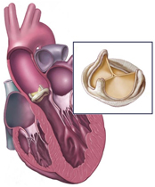
Aortic Valve Stenosis
Symptoms
Aortic stenosis may not produce immediate symptoms or signs. The first sign is usually an abnormal heart sound, or heart murmur, which may develop months or even decades before other signs and symptoms. Aortic stenosis ranges from mild to severe.
As the valve narrows, more signs and symptoms develop and can include : -
Chest pain (angina) or tightness
Feeling faint or fainting with physical exertion (exercise)
Dizziness
Fatigue, especially during times of increased activity
Shortness of breath, especially with physical exertion
Heart palpitations - sensations of a rapid, fluttering heartbeat
Swollen ankles and feet
Causes
Aortic valve stenosis obstructs the way blood normally flows through the heart
Causes may be : -
Calcium buildup on the valve
With age, heart valves may accumulate deposits of calcium (aortic valve calcification). Calcium is a mineral found in your blood. As blood repeatedly flows over the aortic valve, deposits of calcium can accumulate on the valve's leaflets. These deposits may never cause any problems. However, in some people - particularly those with a bicuspid aortic valve - calcium deposits result in stiffening of the valve leaflets. This stiffening narrows the aortic valve. This cause of aortic stenosis is most common in people older than 60, and symptoms often don't appear until age 70 or 80.
Rheumatic fever
This complication of strep throat was once a common childhood illness in the United States. Rheumatic fever may result in scar tissue forming on the aortic valve. Scar tissue can narrow the aortic valve and lead to aortic stenosis. Scar tissue can also create a rough surface on which calcium deposits can collect, contributing to aortic stenosis later in life. Rheumatic fever may damage more than one heart valve, and in more than one way. A damaged heart valve may not open fully or close fully - or both. Many older adults in the United States were exposed to rheumatic fever as children. Rheumatic fever is still prevalent in underdeveloped countries.
Congenital heart defect
Rarely, some babies are born with an already narrowed aortic valve. Others are born with an aortic valve that has only two flaps (leaflets) - not three. Known as a bicuspid aortic valve, this deformity may not cause problems until adulthood, when the valve may begin to narrow or leak and may need to be repair or replacement. Having a bicuspid aortic valve requires regular evaluation by a physician to watch for signs of valve malfunction. Parents of a newborn, infant or child with aortic stenosis may have many questions and concerns. In most cases, physicians don't know why a heart valve fails to develop properly, and it isn't something the parents could have prevented.
Aortic Regurgitation
Symptoms
In most cases, aortic regurgitation develops gradually over decades. The heart compensates for the problem. No signs or symptoms may appear for many years. Most people are unaware they have this condition.
However, as aortic regurgitation progresses, signs and symptoms usually appear and may include : -
Fatigue and weakness, especially during physical exertion
Shortness of breath, especially with physical exertion or when lying down
Chest pain, discomfort or tightness, often increasing during physical exertion
Fainting
Rapid or irregular pulse
Fluttering heart beat
Swollen ankles and feet
Aortic valve regurgitation that occurs suddenly is a medical emergency that requires in-hospital treatment by one or more specialists.
Causes
Any condition that damages the aortic valve can cause regurgitation.
Causes of aortic regurgitation may be : -
Rheumatic fever
This complication of strep throat was once a common childhood illness in the United States. It can damage the aortic valve, leading to aortic regurgitation later in life, and may damage more than one heart valve and in more than one way. A damaged heart valve may not open fully or close fully - or both. Many older adults in the United States were exposed to rheumatic fever as children. Rheumatic fever is still prevalent in underdeveloped countries.
Deterioration of the valve with age
The aortic valve opens and shuts tens of thousands of times a day, every day of a person's life. Aortic regurgitation may result from age-related wear and tear on the valve. We Care partner hospitals surgeons has extensive experience successfully treating elderly patients who have aortic regurgitation.
Endocarditis
The aortic valve may be damaged by endocarditis - an infection inside the heart that can involve the heart valves. Read more about endocarditis prevention.
Congenital heart defect
Some infants are born with an aortic valve that has one leaflet (unicuspid valve) or two leaflets (bicuspid valve) rather than the normal three leaflets. This puts the child at risk of developing aortic regurgitation at some point in their life.
Other causes
Other, rarer conditions can damage the aortic valve and lead to aortic regurgitation, including : -
Marfan syndrome, a disease of connective tissue
Ankylosing spondylitis, a spine disorder
Reactive arthritis, a rare form of arthritis affecting the eyes and joints
Syphilis, a sexually transmitted disease
Damage to the aorta near the aortic valve, such as damage from trauma to the chest or a tear in the aorta, can cause regurgitation
Diagnosis
At We Care partner hospital, diagnosis begins with a complete physical examination by a medical team that specializes in heart care. Patients will be asked about their general health, including signs and symptoms, and a history of heart disease in their family.
Various tests can help diagnose the type of heart valve problem, the possible cause of a heart valve defect, determine how serious the problem is and whether the aortic valve needs to be surgically repaired or replaced.
Diagnostic tests may include : -
Chest X-ray
An X-ray image of the chest allows the physician to study the size and shape of the heart and determine whether the heart's left ventricle (lower left chamber) is enlarged - a possible sign of a damaged aortic valve. A chest X-ray can also reveal calcium deposits on the aortic valve. In addition, a chest X-ray helps the physician check the condition of the lungs. Aortic stenosis may lead to blood and fluid backing up in the lungs, which causes congestion visible on an X-ray.
Electrocardiogram (ECG)
In this test, patches with wires (electrodes) are attached to the patient's skin to measure the electrical impulses given off by the heart. Impulses are recorded as waves displayed on a monitor or printed on paper. An ECG can provide clues about whether the heart's left ventricle is thickened or enlarged, which can occur with aortic stenosis.
Echocardiogram (Doppler Echocardiogram)
This test uses sound waves to produce images of the patient's heart. Through a wandlike device (transducer) held on the patient's chest, sound waves bounce off the heart and are reflected back through the chest wall and processed to produce video images of the heart and a close look at the aortic valve. A Doppler echocardiogram may be used to measure the volume of blood flowing backward through the aortic valve. This volume is expressed in cubic centimeters (cc) per heart beat.
Transesophageal Echocardiogram
Similar to a "regular" echocardiogram, this test uses a tiny transducer (sound device) on a tube inserted down the esophagus (part of the digestive tract that runs from the throat to the stomach). Because the esophagus lies close to the heart, having the transducer placed there provides an even more detailed image of the aortic valve and blood flow through the valve.
Exercise Tests
Different types of exercise tests help measure the patient's tolerance for activity and check the heart's response to physical exertion (exercise).
Cardiac Catheterization
In this procedure, a thin tube (catheter) is inserted in a blood vessel in the patient's arm or groin and threaded up to the heart. The catheter is used to deliver dye into the heart chambers and heart blood vessels. The dye, appearing on X-ray images as it moves through the heart, gives physicians detailed information about the heart and heart valves.
This test helps show blockage in arteries to the heart that can coexist with aortic stenosis and may need surgical treatment at the same time as the aortic stenosis.
Diagnostic Imaging Scans
To provide more detailed, three-dimensional images of the heart, a patient may have a scan using:
Computed tomography (CT) : - A CT scan uses a series of X-rays to create a detailed image.
Magnetic Resonance Imaging (MRI) : - An MRI uses powerful magnets and radio waves to create a detailed image.
Positron Emission Tomography (PET) : - A PET scan involves injecting the body with a small amount of radioactive glucose (tracer), which can be tracked by a special camera (positron) to provide detailed images.
We Care partner hospital has other tests that use nuclear medicine (radioactive materials) for detailed imaging and advanced analysis of the heart and heart valves.
Surgery
Many factors help determine the appropriate surgery to treat heart valve disease, including a patient's age and general health, the extent of damage to the valves, the type of valve, and the patient's preference. Many of these procedures can be done using minimally invasive heart surgery.
Surgery for Heart Valve Repair or Replacement : -
Stenosis (valve narrowing) may be corrected by cutting, separating or reconstructing the valve leaflets, or other valve components, to widen the valve opening. Most patients with stenosed valves require valve replacement.
Regurgitation (valve leakage) may be corrected by replacing or shortening the supporting valve structures to allow the valve to close tightly, or by inserting a prosthetic ring to reshape a deformed valve. Valve flaps (leaflets) may also be modified to stop blood from flowing backwards.
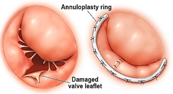
Annuloplasty : -
Annuloplasty is one type of heart valve surgery.
Annuloplasty describes surgical techniques performed on the valve's annulus, the ring of tissue that supports the valve leaflets. Purse-string sutures are sewn around the ring to make the opening smaller which helps the leaflets meet again when the valve closes.
Sometimes when repairing the annulus, it is necessary to implant an annuloplasty ring where the leaflets meet the fibrous annulus to downsize an abnormally enlarged valve opening so the leaflets can come together properly.
Repair of structural support : -
Repair of structural support replaces or shortens the cords that support the valves (chordae tendineae and papillary muscles). When the cords and muscles are the right length, the valve leaflet edges meet and eliminate the leak.
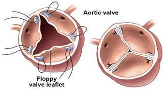
Valve repair (Valvuloplasty) : -
Valvuloplasty is one type of heart valve surgery.
Valve repair may be done when a valve leaflet is floppy and prolapses. The procedure involves surgically separating, cutting out a section, or pleating a valve leaflet.
Valve replacement : -
Valve replacement involves removal of the defective valve and replacing it with an artificial (prosthetic) valve by sewing it to the annulus of the natural valve. Approximately 95 percent of all valve replacements are performed for mitral or aortic valves.
Treatment
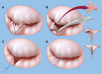
Heart Valve Repair : -
Heart specialists and surgeons agree that whenever possible a heart valve should be repaired instead of replaced. Heart valve repair leaves patients with their normally functioning tissue, which resists infection and does not require a lifetime of blood-thinning medication. Patients who have valve repair generally have a longer life expectancy.
Part of a prolapsing mitral valve is removed to allow the valve to fully close and stop leaking.
Valve repair may be performed to separate fused valve leaflets, sew torn leaflets or reshape parts of the valve. However, repair is not possible for severely damaged valves such as those affected by calcium deposits or rheumatic disease.\
Cardiovascular surgeons at We Care partner hospitals have pioneered, studied and taught cutting-edge valve repair techniques since the beginning of cardiac surgery. In some cases, they can now offer the same proven techniques through smaller, incisions using minimally invasive heart surgery.
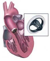
Heart Valve Replacement : -
Heart valves that are severely damaged must be replaced. Valve replacement is most often used to treat aortic valves and badly damaged mitral valves, but it can be used to treat any valve disease that is life threatening.
Two kinds of replacement valves are available : -
1 ) Mechanical valves
Biological valves are made of animal or human tissue.
Mechanical valves are made of synthetic materials. They are reliable, and last a long time. Because blood tends to stick to mechanical valves and create blood clots, patients with these valves will need to take blood-thinning medicines (anticoagulants).
2 ) Biological valves
Biological valves are made from animal tissue (called a xenograft) or taken from the human tissue of a donated heart (called an allograft or homograft). Sometimes, a patient's own tissue can be used for valve replacement (called an autograft/Ross procedure).
Patients with biological valves usually do not need to take blood-thinning medication. These valves are not as durable as mechanical valves, however, and they may need to be replaced. Biological valves are used most often in elderly patients.
The choice of valve should be made by the patient and the physician, taking the following into consideration : -
age
other medical conditions
the patient's preferences with regard to medications and the possible need for reoperation
potential pregnancy in women of childbearing age
lifestyle
The list of of world class heart hospitals in India is as follows : -
For more information, medical assessment and medical quote
send your detailed medical history and medical reports
as email attachment to
Email : - info@wecareindia.com
Call: +91 9029304141 (10 am. To 8 pm. IST)
(Only for international patients seeking treatment in India)
For a detailed evaluation send patient’s medical reports / X rays / doctors notes to info@wecareindia.com
Patient Storys
Successful heart surgery at We Care India partner hospital allows Robert Clarke to live a normal life despite a rare genetic disorder We Care india helped Robert find best super specialised surgeon for his rare conditions.